Search Thermo Fisher Scientific
Disclaimer

Clicking the images or links will redirect you to a website hosted by BenchSci that provides third-party scientific content. Neither the content nor the BenchSci technology and processes for selection have been evaluated by us; we are providing them as-is and without warranty of any kind, including for use or application of the Thermo Fisher Scientific products presented.
Invitrogen
CD366 (TIM3) Monoclonal Antibody (8B.2C12), Functional Grade, eBioscience™
 Antibody in Flow Cytometry (Flow)")
FIGURE: 1 / 8
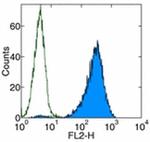
CD366 (TIM3) Antibody (16-5871-85) in Flow
Staining of CD366 transfected cells with Rat IgG1 K Isotype Control Functional Grade Purified (Product # 16-4301-81) (open histogram) or Anti-Mouse CD366 (TIM-3) Functional Grade Purified (filled histogram) followed by Anti-Rat IgG Biotin (Product # 13-4813-85) and Streptavidin PE (Product # 12-4317-87).Total viable cells were used for analysis.


 Antibody (16-5871-85) in Flow")
 Antibody (16-5871-85) in WB, IP")
 Antibody (16-5871-85) in Flow")
 Antibody (16-5871-85) in Flow")
 Antibody (16-5871-85) in Flow")
 Antibody (16-5871-85) in Flow")
 Antibody (16-5871-85) in Flow")
 Antibody (16-5871-85) in Neu")
Product Details
16-5871-85
Applications
Tested Dilution
Publications
Product Specifications
Species Reactivity
Mouse
Published species
Mouse
Host/Isotype
Rat
/ IgG1, kappa
Recommended Isotype Control
Class
Monoclonal
Type
Antibody
Clone
8B.2C12
Conjugate
Functional Grade
Form
Liquid
Concentration
1 mg/mL
Purification
Affinity chromatography
Storage buffer
PBS, pH 7.2
Contains
no preservative
Storage conditions
4° C
Shipping conditions
Wet ice
RRID
AB_469105
Product Specific Information
Description: The 8B.2C12 monoclonal antibody reacts with mouse CD366 (TIM3), a Th1-specific cell surface protein. CD366 is a type I transmembrane protein and contains an immunoglobulin and a mucin-like domain in its extracellular portion and a tyrosine phosphorylation motif in its cytoplasmic portion. CD366 is expressed selectively by differentiated CD4+ Th1 and CD8+ Tc1, but is absent on Th2 and Tc2. Other hematopoietic cell types, including naive T cells, B cells, macrophages and dendritic cells, do not express CD366, at least at the protein level. Expression of CD366 is upregulated at a late stage of T cell differentiation on Th1 cells after 3 rounds of in vitro polarization suggesting a role for this molecule in the transport or effector function of Th1 cells rather than a contribution to T cell differentiation. In an experimental autoimmune encephalomyelitis (EAE) model, CD366 was shown to be expressed on most CD4+ and CD8+ T cells in the central nervous system at the onset of clinical signs of disease, while less than 2% of CD4+ cells in the periphery expressed CD366 after immunization. In this model, in vivo administration of 8B.2C12 resulted in a hyperacute and atypical disease phenotype. It is postulated that the engagement of CD366 during T cell activation results in the expansion and activation of macrophages and increased severity of autoimmune disease. The Tim gene family may have an important role in the regulation of autoimmunity and allergies.
The 8B.2C12 antibody binds to the BALB/c allele of CD366 while reactivity to the C57Bl/6 allele is significantly weaker.
Applications Reported: 8B.2C12 has been reported for use in flow cytometric analysis and functional studies.
Applications Tested: The 8B.2C12 antibody has been tested by flow cytometric analysis of mouse splenocytes and mouse Tim-3 transfected cells and can be used at less than or equal to 0.5 µg per test. A test is defined as the amount (µg) of antibody that will stain a cell sample in a final volume of 100 µL. Cell number should be determined empirically but can range from 10^5 to 10^8 cells/test. It is recommended that the antibody be carefully titrated for optimal performance in the assay of interest.
Storage and handling: Use in a sterile environment.
Filtration: 0.2 µm post-manufacturing filtered.
Purity: Greater than 90%, as determined by SDS-PAGE.
Endotoxin Level: Less than 0.001 ng/µg antibody, as determined by LAL assay.
Aggregation: Less than 10%, as determined by HPLC.
Target Information
TIM3 (Hepatitis A virus cellular receptor 2, HAVCR2, T-cell immunoglobulin, mucin-dmain containing-3) is a 281 amino acid long, Type-1 Th1- specific cell surface glycoprotein expressed on terminally differentiated CD4+Th1 and CD8+Tc1 cells. TIM3 consists of an IgV-like domain, a mucin-like domain in the extracellular region, and a conserved Tyrosine phosphorylation motif in the cytoplasmic region. TIM3 is involved in macrophage activation and induction of autoimmune diseases. Further, TIM3 down-regulates aggressive Th1-mediated immune responses and facilitates in the development of immune tolerance. Pathological significance of TIM3 has been attributed to Experimental autoimmune encephalomyelitis (EAE), a Th-1 dependent autoimmune disease, and also enhances the severity of experimental autoimmune encephalomyelitis in mice.
For Research Use Only. Not for use in diagnostic procedures. Not for resale without express authorization.
Bioinformatics
Protein Aliases: CD366; FLJ14428; HAVcr-2; Hepatitis A virus cellular receptor 2 homolog; sCD366; soluble CD366; soluble TIM 3; T-cell immunoglobulin and mucin domain containing 3; T-cell immunoglobulin and mucin domain-containing protein 3; T-cell immunoglobulin mucin receptor 3; T-cell membrane protein 3; TIM-3; TIMD-3
Gene Aliases: Havcr2; TIM-3; Tim3; Timd3
UniProt ID: (Mouse) Q8VIM0
Entrez Gene ID: (Mouse) 171285
Molecular Function:
![]() immunoglobulin receptor superfamily
immunoglobulin receptor superfamily

Performance Guarantee
If an Invitrogen™ antibody doesn't perform as described on our website or datasheet,we'll replace the product at no cost to you, or provide you with a credit for a future purchase.*
Learn more
We're here to help
Get expert recommendations for common problems or connect directly with an on staff expert for technical assistance related to applications, equipment and general product use.
Contact tech support