Search Thermo Fisher Scientific
Disclaimer

Clicking the images or links will redirect you to a website hosted by BenchSci that provides third-party scientific content. Neither the content nor the BenchSci technology and processes for selection have been evaluated by us; we are providing them as-is and without warranty of any kind, including for use or application of the Thermo Fisher Scientific products presented.
Invitrogen
beta Catenin Monoclonal Antibody (15B8), Alexa Fluor™ 488, eBioscience™
")
FIGURE: 1 / 8
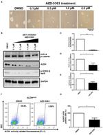
beta Catenin Antibody (53-2567-41) in Flow
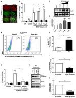
Intracellular staining of the Jurkat cell line using the Foxp3 Staining Buffer Set (Product # 00-5523-00) with Mouse IgG1 K Isotype Control Alexa Fluor® 488 (Product # 53-4714-42) (blue histogram) or Anti-Human/Mouse beta-Catenin Alexa Fluor® 488 (purple histogram).


 in Flow")
 in WB, ICC/IF")
 in ICC/IF, IHC")
 in ICC/IF")
 in WB")
 in WB")
 in WB")
 in Flow")
Product Details
53-2567-41
Applications
Tested Dilution
Publications
Product Specifications
Species Reactivity
Human,
Mouse
Published species
Human
Host/Isotype
Mouse
/ IgG1, kappa
Recommended Isotype Control
Class
Monoclonal
Type
Antibody
Clone
15B8
Conjugate
Alexa Fluor™ 488
Excitation/Emission Max
499/520 nm
View spectra

Form
Liquid
Concentration
5 µL/Test
Purification
Affinity chromatography
Storage buffer
PBS, pH 7.2, with 0.2% BSA
Contains
0.09% sodium azide
Storage conditions
4° C, store in dark, DO NOT FREEZE!
Shipping conditions
Wet ice
RRID
AB_10807094
Product Specific Information
Description: The 15B8 monoclonal antibody reacts with human and mouse beta-catenin, one member of a family of catenins, which are intracellular proteins that interact with cadherins to mediate cellular adhesion. More specifically, beta-catenin binds to the cytoplasmic tail of E-cadherin. In addition, this molecule is a component of the canonical Wnt signaling pathway. In the absence of Wnt binding its receptor, beta-catenin is phosphorylated and resides in the cytoplasm where it is eventually targeted for degradation by ubiquitination. Upon Wnt binding, beta-catenin becomes dephosphorylated, translocates to the nucleus, and modulates gene expression in partnership with the transcription factors T cell factor (TCF) and lymphocyte enhancer binding factor (LEF). Expression of beta-catenin is found in a wide variety of non-immune and immune tissues, including thymocytes and T and B lymphocytes. The Wnt and beta-catenin signaling pathway has been demonstrated to play a crucial role in the development of T, B, and hematopoietic stem cells.
Applications Reported: This 15B8 antibody has been reported for use in intracellular staining followed by flow cytometric analysis.
Applications Tested: This 15B8 antibody has been pre-titrated and tested by intracellular staining and flow cytometric analysis of Jurkat cell line using the Foxp3/Transcription Factor Staining Buffer Set (cat. 00-5523). This can be used at 5 µL (0.25 µg) per test. A test is defined as the amount (µg) of antibody that will stain a cell sample in a final volume of 100 µL. Cell number should be determined empirically but can range from 10^5 to 10^8 cells/test.
Excitation: 488 nm; Emission: 519 nm; Laser: Blue Laser.
Filtration: 0.2 µm post-manufacturing filtered.
Target Information
Beta-catenin, an adherens junction (AJ) protein, was originally identified as a component of cell-cell adhesion structures. AJs are necessary for the creation and maintenance of epithelial cell layers by regulating cell growth and adhesion between cells. Beta-catenin interacts with the cytoplasmic domain of E-cadherin and links E-cadherin to alpha-catenin, which in turn mediates anchorage of the E-cadherin complex to the cortical actin cytoskeleton. Studies show that Beta-catenin also binds to another cytoskeletal complex containing the adenomatous polyposis coli protein and microtubules, and interacts with several signaling pathways that include tyrosine kinases, phosphatases and Wnt/Wingless. The interplay between beta-catenin, cytoskeletal complexes and signaling pathways may regulate morphogenesis. Beta-catenin is expressed in several hair follicle cell types, basal and peripheral matrix cells, and cells of the outer and inner root sheats. A pathological role of beta-catenin has been identified in pilomatrixoma (PTR), medulloblastoma (MDB), colorectal cancer (CRC), ovarian cancer, and tumor development. In the nucleus, beta-catenin serves to co activate a family of Lef/Tcf transcription factors that stimulate transcription of target genes including those encoding cyclin D and c-myc that promote cell proliferation. The influence on cell proliferation is the molecular basis for the role of beta-catenin in tumorgenesis, specifically, solid tumors of the breast, colon, liver, lung, gastric, prostate, and skin.
For Research Use Only. Not for use in diagnostic procedures. Not for resale without express authorization.
Flow Cytometry
Panel Builder
How to use the Panel Builder
Watch the video to learn how to use the Invitrogen Flow Cytometry Panel Builder to build your next flow cytometry panel in 5 easy steps.
Bioinformatics
Protein Aliases: Bcatenin; beta 1 88kDa; Beta-catenin; Betacatenin; C20orf33; Cadherin associated protein; catenin; catenin (cadherin-associated protein), beta 1; catenin (cadherin-associated protein), beta 1, 88kDa; Catenin b 1; Catenin b1; Catenin beta-1; Catenin beta1; Catenin β 1; Catenin β1; CTNB1; dJ633O20.1; DKFZp686D02253; FLJ25606; FLJ37923; NYD-SP19; OTTHUMP00000209289; P14L; PP8304; RP5-1118M15.1; β catenin; βcatenin
Gene Aliases: armadillo; Bfc; Catnb; CTNNB; CTNNB1; Mesc; MRD19; OK/SW-cl.35; PRO2286
UniProt ID: (Human) P35222, (Mouse) Q02248
Entrez Gene ID: (Human) 1499, (Mouse) 12387
negative regulation of transcription from RNA polymerase II promoter
embryonic axis specification
cell morphogenesis involved in differentiation
skeletal system development
patterning of blood vessels
vasculogenesis
branching involved in ureteric bud morphogenesis
in utero embryonic development
gastrulation with mouth forming second
endoderm formation
cell fate specification
cell fate determination
endodermal cell fate commitment
neuron migration
kidney development
neural plate development
vasculature development
positive regulation of neuroblast proliferation
positive regulation of mesenchymal cell proliferation
lens morphogenesis in camera-type eye
ventricular compact myocardium morphogenesis
regulation of secondary heart field cardioblast proliferation
mesenchymal to epithelial transition involved in metanephros morphogenesis
metanephros morphogenesis
transcription, DNA-templated
regulation of transcription, DNA-templated
regulation of transcription from RNA polymerase II promoter
cell adhesion
cell-matrix adhesion
chemical synaptic transmission
ectoderm development
nervous system development
glial cell fate determination
heart development
cell proliferation
positive regulation of cell proliferation
negative regulation of cell proliferation
anterior/posterior axis specification
dorsal/ventral axis specification
dorsal/ventral pattern formation
proximal/distal pattern formation
cellular process
regulation of gene expression
positive regulation of gene expression
positive regulation of epithelial to mesenchymal transition
positive regulation of heparan sulfate proteoglycan biosynthetic process
Schwann cell proliferation
Wnt signaling pathway
morphogenesis of embryonic epithelium
single organismal cell-cell adhesion
stem cell population maintenance
layer formation in cerebral cortex
central nervous system vasculogenesis
hair cycle process
hemopoiesis
cell differentiation
T cell differentiation
osteoclast differentiation
lung development
male genitalia development
regulation of epithelial cell differentiation
positive regulation of epithelial cell differentiation
forebrain development
regulation of centriole-centriole cohesion
pancreas development
hair follicle morphogenesis
regulation of myelination
positive regulation of telomere maintenance via telomerase
negative regulation of chondrocyte differentiation
response to estradiol
T cell differentiation in thymus
negative regulation of protein sumoylation
response to cytokine
adherens junction organization
adherens junction assembly
protein localization to cell surface
cellular protein localization
embryonic heart tube development
genitalia morphogenesis
embryonic forelimb morphogenesis
embryonic hindlimb morphogenesis
embryonic skeletal limb joint morphogenesis
regulation of cell proliferation
regulation of T cell proliferation
odontogenesis of dentin-containing tooth
regulation of cell fate specification
embryonic digit morphogenesis
regulation of apoptotic process
positive regulation of apoptotic process
positive regulation of I-kappaB kinase/NF-kappaB signaling
positive regulation of MAPK cascade
skin development
response to estrogen
canonical Wnt signaling pathway involved in positive regulation of epithelial to mesenchymal transition
canonical Wnt signaling pathway involved in negative regulation of apoptotic process
bone resorption
regulation of cell differentiation
negative regulation of cell differentiation
positive regulation of endothelial cell differentiation
regulation of osteoblast differentiation
positive regulation of osteoblast differentiation
regulation of osteoclast differentiation
negative regulation of osteoclast differentiation
positive regulation of fibroblast growth factor receptor signaling pathway
negative regulation of transcription, DNA-templated
positive regulation of transcription, DNA-templated
positive regulation of transcription from RNA polymerase II promoter
negative regulation of mitotic cell cycle, embryonic
chromatin-mediated maintenance of transcription
cell maturation
synaptic vesicle transport
animal organ development
thymus development
oocyte development
embryonic foregut morphogenesis
regulation of smooth muscle cell proliferation
negative regulation of oligodendrocyte differentiation
synapse organization
smooth muscle cell differentiation
protein heterooligomerization
positive regulation of telomerase activity
cardiac muscle cell proliferation
oviduct development
canonical Wnt signaling pathway
limb development
trachea morphogenesis
trachea formation
epithelial tube branching involved in lung morphogenesis
lung cell differentiation
lung-associated mesenchyme development
mesenchyme development
lung induction
epithelial cell differentiation involved in prostate gland development
positive regulation of epithelial cell proliferation involved in prostate gland development
hair follicle placode formation
mesenchymal cell proliferation involved in lung development
cardiac vascular smooth muscle cell differentiation
coronary artery morphogenesis
epicardium-derived cardiac vascular smooth muscle cell differentiation
positive regulation of branching involved in lung morphogenesis
endothelial tube morphogenesis
fungiform papilla formation
canonical Wnt signaling pathway involved in positive regulation of cardiac outflow tract cell proliferation
sympathetic ganglion development
regulation of centromeric sister chromatid cohesion
cellular response to mechanical stimulus
cellular response to growth factor stimulus
cellular response to organic cyclic compound
cellular response to indole-3-methanol
renal system development
renal vesicle formation
renal inner medulla development
renal outer medulla development
nephron tubule formation
mesenchyme morphogenesis
regulation of nephron tubule epithelial cell differentiation
regulation of calcium ion import
negative regulation of neuron death
cellular response to insulin-like growth factor stimulus
regulation of protein localization to cell surface
positive regulation of determination of dorsal identity
negative regulation of apoptotic signaling pathway
epithelial to mesenchymal transition
liver development
negative regulation of mesenchymal to epithelial transition involved in metanephros morphogenesis
Wnt signaling pathway, calcium modulating pathway
midgut development
response to activity
androgen receptor signaling pathway
hindbrain development
positive regulation of type I interferon production
hair cell differentiation
catenin import into nucleus
response to drug
proteasome-mediated ubiquitin-dependent protein catabolic process
positive regulation of neuron apoptotic process
cellular response to fibroblast growth factor stimulus
myoblast differentiation
regulation of angiogenesis
response to cadmium ion
regulation of fibroblast proliferation
positive regulation of skeletal muscle tissue development
embryonic cranial skeleton morphogenesis
regulation of neurogenesis
positive regulation of sequence-specific DNA binding transcription factor activity
positive regulation of muscle cell differentiation
positive regulation of histone H3-K4 methylation
cranial ganglion development
cellular response to lithium ion
positive regulation of chromatin-mediated maintenance of transcription
regulation of euchromatin binding
regulation of core promoter binding
beta-catenin-TCF complex assembly
beta-catenin destruction complex disassembly
midbrain dopaminergic neuron differentiation
canonical Wnt signaling pathway involved in midbrain dopaminergic neuron differentiation
embryonic brain development
dorsal root ganglion development
positive regulation of DNA-templated transcription, initiation
RNA polymerase II transcription factor binding
RNA polymerase II activating transcription factor binding
double-stranded DNA binding
transcription factor activity, sequence-specific DNA binding
transcription coactivator activity
signal transducer activity
protein binding
protein C-terminus binding
transcription factor binding
enzyme binding
kinase binding
protein kinase binding
protein phosphatase binding
estrogen receptor binding
protein complex binding
ionotropic glutamate receptor binding
nuclear hormone receptor binding
transcription regulatory region DNA binding
ion channel binding
alpha-catenin binding
cadherin binding
SMAD binding
androgen receptor binding
nitric-oxide synthase binding
I-SMAD binding
repressing transcription factor binding
cadherin binding involved in cell-cell adhesion
euchromatin binding
DNA binding
chromatin binding
R-SMAD binding

Performance Guarantee
If an Invitrogen™ antibody doesn't perform as described on our website or datasheet,we'll replace the product at no cost to you, or provide you with a credit for a future purchase.*
Learn more
We're here to help
Get expert recommendations for common problems or connect directly with an on staff expert for technical assistance related to applications, equipment and general product use.
Contact tech support