Search Thermo Fisher Scientific
Disclaimer

Clicking the images or links will redirect you to a website hosted by BenchSci that provides third-party scientific content. Neither the content nor the BenchSci technology and processes for selection have been evaluated by us; we are providing them as-is and without warranty of any kind, including for use or application of the Thermo Fisher Scientific products presented.
Invitrogen
HLA-DR Monoclonal Antibody (LN3), APC-eFluor™ 780, eBioscience™
Advanced Verification
This Antibody was verified by Cell treatment to ensure that the antibody binds to the antigen stated.
")
FIGURE: 1 / 29
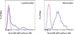
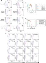
HLA-DR Antibody (47-9956-41) in Flow
Normal human peripheral blood cells were stained with Mouse IgG2b kappa Isotype Control, APC-eFluor 780 (Product # 47-4732-80) (blue histogram) or HLA-DR Monoclonal Antibody, APC-eFluor 780 (purple histogram). Cells in the lymphocyte gate (left) or monocyte gate (right) were used for analysis.


 in Flow")
 in Flow")
 in Flow")
 in Flow")
 in Flow")
 in Flow")
 in Flow")
 in Flow")
 in Flow")
 in Flow")
 in Flow")
 in Flow")
 in Flow")
 in Flow")
 in Flow")
 in Flow")
 in Flow")
 in Flow")
 in Flow")
 in Flow")
 in Flow")
 in Flow")
 in Flow")
 in Flow")
 in Flow")
 in Flow")
 in Flow")
")
")
Product Details
47-9956-41
Applications
Tested Dilution
Publications
Product Specifications
Species Reactivity
Human
Published species
Human
Host/Isotype
Mouse
/ IgG2b, kappa
Recommended Isotype Control
Class
Monoclonal
Type
Antibody
Clone
LN3
Conjugate
APC-eFluor™ 780
Excitation/Emission Max
756/785 nm
View spectra

Form
Liquid
Concentration
5 µL/Test
Purification
Affinity chromatography
Storage buffer
PBS, pH 7.2, with 0.2% BSA
Contains
0.09% sodium azide
Storage conditions
4° C, store in dark, DO NOT FREEZE!
Shipping conditions
Wet ice
RRID
AB_1963604
Product Specific Information
Description: The LN3 mAb reacts with the human major histocompatibility complex (MHC) class II, HLA-DR. HLA-DR is expressed on the surface of human antigen presenting cells (APC) including B cells, monocytes, macrophages, DCs, and activated T cells. HLA-DR is a heterodimeric transmembrane protein composed of alpha and beta subunits and plays an important role in the presentation of peptides to CD4^+ T lymphocytes.
Applications Reported: This LN3 antibody has been reported for use in flow cytometric analysis.
Applications Tested: This LN3 antibody has been pre-titrated and tested by flow cytometric analysis of normal human peripheral blood cells. This can be used at 5 µL (0.03 µg) per test. A test is defined as the amount (µg) of antibody that will stain a cell sample in a final volume of 100 µL. Cell number should be determined empirically but can range from 10^5 to 10^8 cells/test.
APC-eFluor 780 emits at 780 nm and is excited with the Red laser (633 nm). Please make sure that your instrument is capable of detecting this fluorochome.
Light sensitivity: This tandem is sensitive to photo-induced oxidation. Please protect this vial and stained samples from light.
Fixation: Samples can be stored in IC Fixation Buffer (cat. 00-8222) (100 µL cell sample + 100 µL IC Fixation Buffer) or 1-step Fix/Lyse Solution (cat. 00-5333) for up to 3 days in the dark at 4°C with minimal impact on brightness and FRET efficiency/compensation. Some generalizations regarding fluorophore performance after fixation can be made, but clone specific performance should be determined empirically.
Excitation: 633-647 nm; Emission: 780 nm; Laser: Red Laser.
Filtration: 0.2 µm post-manufacturing filtered.
Target Information
HLA-DR, like other MHC class II molecules, is a transmembrane glycoprotein composed of a 36 kDa alpha chain (DRA) and 27 kDa beta chain (DRB). The alpha chain gene contains 5 exons. Exon 1 encodes the leader peptide, exons 2 and 3 encode the two extracellular domains, and exon 4 encodes the transmembrane domain and the cytoplasmic tail. DRA does not have polymorphisms in the peptide binding part and acts as the sole alpha chain for DRB1, DRB3, DRB4 and DRB5. Within the DR molecule the beta chain contains all the polymorphisms specifying the peptide binding specificities. Hundreds of DRB1 alleles have been described and typing for these polymorphisms is routinely done for bone marrow and kidney transplantation. HLA-DR is expressed primarily on antigen presenting cells such as B lymphocytes, monocytes, macrophages, thymic epithelial cells and activated T lymphocytes. Three loci, DR, DQ and DP, encode the major expressed products of the human class II region. The human MHC class II molecules bind intracellularly processed peptides, present them to T-helper cells, and have a critical role in the initiation of the immune response.
HLA and MHC antibodies play a significant role in Immunopeptidomics, facilitating the identification and characterization of neoantigens through high-performance liquid chromatography coupled to tandem Mass Spectrometry.
For Research Use Only. Not for use in diagnostic procedures. Not for resale without express authorization.
Flow Cytometry
Panel Builder
How to use the Panel Builder
Watch the video to learn how to use the Invitrogen Flow Cytometry Panel Builder to build your next flow cytometry panel in 5 easy steps.
Bioinformatics
Protein Aliases: CD74; CD74 antigen; CD74 antigen (invariant polypeptide of major histocompatibility complex, class II antigen-associated); CD74 molecule, major histocompatibility complex, class II invariant chain; Class-II-associated invariant chain peptide; CLIP; DASS-397D15.1; DR beta-5; DR-9; DR2-beta-2; DR7; DR9; DRB1 transplantation antigen; Dw2; DW2.2/DR2.2; FLJ51114; FLJ75017; FLJ76359; gamma chain of class II antigens; histocompatibility antigen HLA-DR alpha; HLA class II histocompatibility antigen gamma chain; HLA class II histocompatibility antigen, DR alpha chain; HLA class II histocompatibility antigen, DR beta 3 chain; HLA class II histocompatibility antigen, DR beta 4 chain; HLA class II histocompatibility antigen, DR beta 5 chain; HLA class II histocompatibility antigen, DR-1 beta chain; HLA class II histocompatibility antigen, DR-5 beta chain; HLA class II histocompatibility antigen, DRB1 beta chain; HLA class II histocompatibility antigen, DRB1-7 beta chain; HLA class II histocompatibility antigen, DRB1-9 beta chain; HLA-DR antigens-associated invariant chain; HLA-DR-gamma; HLA-DRB1; HLA-DRB5; human leucocyte antigen DRB1; human leucocyte antigen DRB3; human leucocyte antigen DRB4; human leucocyte antigen DRB5; Human leukocyte antigen DRB1; Ia antigen-associated invariant chain; Ia-associated invariant chain; Ii; leukocyte antigen; leukocyte antigen class II; lymphocyte antigen DRB1; MHC cell surface glycoprotein; MHC class I antigen; MHC Class II antigen; MHC class II antigen DR beta 3 chain; MHC class II antigen DRA; MHC class II antigen DRB1*9; MHC class II antigen DRB3; MHC class II antigen DRB4; MHC class II antigen DRB5; MHC class II antigen HLA-DR-beta; MHC class II HLA beta chain; MHC class II HLA-DR beta 1 chain; MHC class II HLA-DR beta 3 chain; MHC class II HLA-DR-beta cell surface glycoprotein; MHC class II HLA-DRw10-beta; MHC class2 antigen; MHC HLA DR-beta chain; MHC HLA-DR gamma chain; MHC HLA-DR-beta cell surface glycoprotein; MHC HLA-DR-beta chain; p33; XXbac-BPG161M6.1
Gene Aliases: CD74; DHLAG; DR-4; DR4; DRB1; DRB4; DRw10; HLA-DR1B; HLA-DR3B; HLA-DR4B; HLA-DRA; HLA-DRA1; HLA-DRB; HLA-DRB1; HLA-DRB3; HLA-DRB4; HLA-DRB5; HLADG; Ia-GAMMA; II; MLRW; SS1
UniProt ID: (Human) P04233, (Human) P01903, (Human) Q4PRC3, (Human) P79483, (Human) Q6TLK6, (Human) Q30154
Entrez Gene ID: (Human) 972, (Human) 3122, (Human) 3123, (Human) 3125, (Human) 3126, (Human) 3127
Molecular Function:
![]() major histocompatibility complex protein
major histocompatibility complex protein
![]() scaffold/adaptor protein
scaffold/adaptor protein
activation of MAPK activity
prostaglandin biosynthetic process
positive regulation of cytokine-mediated signaling pathway
positive regulation of dendritic cell antigen processing and presentation
negative regulation of peptide secretion
positive regulation of type 2 immune response
negative regulation of mature B cell apoptotic process
protein complex assembly
intracellular protein transport
defense response
signal transduction
cell proliferation
immunoglobulin mediated immune response
antigen processing and presentation of endogenous antigen
antigen processing and presentation of exogenous peptide antigen via MHC class II
positive regulation of B cell proliferation
macrophage migration inhibitory factor signaling pathway
regulation of macrophage activation
negative regulation of apoptotic process
negative regulation of DNA damage response, signal transduction by p53 class mediator
T cell selection
positive thymic T cell selection
negative thymic T cell selection
negative regulation of T cell differentiation
positive regulation of T cell differentiation
positive regulation of fibroblast proliferation
positive regulation of peptidyl-tyrosine phosphorylation
leukocyte migration
chaperone mediated protein folding requiring cofactor
positive regulation of macrophage cytokine production
positive regulation of ERK1 and ERK2 cascade
positive regulation of neutrophil chemotaxis
negative regulation of intrinsic apoptotic signaling pathway in response to DNA damage by p53 class mediator
positive regulation of chemokine (C-X-C motif) ligand 2 production
peptide antigen assembly with MHC class II protein complex
antigen processing and presentation of peptide or polysaccharide antigen via MHC class II
polysaccharide assembly with MHC class II protein complex
immune response
viral process
T cell costimulation
T cell receptor signaling pathway
cognition
interferon-gamma-mediated signaling pathway
beta-amyloid binding
cytokine receptor activity
protein binding
cytokine binding
MHC class II protein complex binding
macrophage migration inhibitory factor binding
MHC class II protein binding
MHC class II protein binding, via antigen binding groove
identical protein binding
protein binding involved in protein folding
nitric-oxide synthase binding
MHC class II receptor activity
peptide antigen binding

Performance Guarantee
If an Invitrogen™ antibody doesn't perform as described on our website or datasheet,we'll replace the product at no cost to you, or provide you with a credit for a future purchase.*
Learn more
We're here to help
Get expert recommendations for common problems or connect directly with an on staff expert for technical assistance related to applications, equipment and general product use.
Contact tech support