Search Thermo Fisher Scientific
Disclaimer

Clicking the images or links will redirect you to a website hosted by BenchSci that provides third-party scientific content. Neither the content nor the BenchSci technology and processes for selection have been evaluated by us; we are providing them as-is and without warranty of any kind, including for use or application of the Thermo Fisher Scientific products presented.
Invitrogen
CD39 Monoclonal Antibody (eBioA1 (A1)), FITC, eBioscience™
")
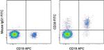
FIGURE: 1 / 15
CD39 Antibody (11-0399-42) in Flow
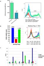
Staining of normal human peripheral blood cells with Anti-Human CD19 APC (Product # 17-0199-42) and Mouse IgG1 kappa Isotype Control FITC (Product # 11-4714-42) (left) or Anti-Human CD39 FITC (right). Cells in the lymphocyte gate were used for analysis.


 in Flow")
 in Flow")
 in Flow")
 in Flow")
 in Flow")
 in Flow")
 in Flow")
 in Flow")
 in Flow")
 in Flow")
 in Flow")
 in Flow")
 in Flow")
 in Flow")
 in IHC")
Product Details
11-0399-42
Applications
Tested Dilution
Publications
Product Specifications
Species Reactivity
Human
Published species
Human,
Rhesus monkey
Host/Isotype
Mouse
/ IgG1, kappa
Recommended Isotype Control
Class
Monoclonal
Type
Antibody
Clone
eBioA1 (A1)
Conjugate
FITC
Excitation/Emission Max
498/517 nm
View spectra

Form
Liquid
Concentration
5 µL/Test
Purification
Affinity chromatography
Storage buffer
PBS, pH 7.2, with 0.2% BSA
Contains
0.09% sodium azide
Storage conditions
4° C, store in dark, DO NOT FREEZE!
Shipping conditions
Wet ice
RRID
AB_11151149
Product Specific Information
Description: The eBioA1 monoclonal antibody reacts with human CD39 also known as ectonucleoside triphosphate diphosphohydrolase 1 (ENTPD1) or NTPDase. CD39 is an integral membrane protein with two transmembrane domains and exists as a homotetramer. It is the most prominent ectoenzyme of the immune system. The function of CD39 is to effectively remove toxic extracellular ATP by converting it to ADP or AMP. CD39 is thought to work together with CD73 to hydrolyze ATP and has been well characterized on Langerhans cells. Expression of CD39 was originally identified on activated lymphocytes. Expression is also found on a subset of T cells, B cells and dendritic cells as well as weak staining on monocytes and granulocytes.
Recently, CD39 and CD73 have been found on regulatory T cells (Treg). Expression of CD39 on Treg may facilitate their entry into inflamed areas where high levels of ATP are present. Expression of CD39 on Foxp3+CD4+ cells ranges from 25-45%.
Applications Reported: This eBioA1 (A1) antibody has been reported for use in flow cytometric analysis.
Applications Tested: This eBioA1 (A1) antibody has been pre-titrated and tested by flow cytometric analysis of normal human peripheral blood cells. This can be used at 5 µL (0.25 µg) per test. A test is defined as the amount (µg) of antibody that will stain a cell sample in a final volume of 100 µL. Cell number should be determined empirically but can range from 10^5 to 10^8 cells/test.
Excitation: 488 nm; Emission: 520 nm; Laser: Blue Laser.
Filtration: 0.2 µm post-manufacturing filtered.
Target Information
CD39 cell surface antigen is a 70-100kD molecule expressed on peripheral blood B cells, monocytes, T cell clones, and also weakly expressed on granulocytes. CD39 has intrinsic ecto-ATPase activity. Expression of CD39 is induced on T cells and increased on B cells as a late activation antigen.
For Research Use Only. Not for use in diagnostic procedures. Not for resale without express authorization.
Flow Cytometry
Panel Builder
How to use the Panel Builder
Watch the video to learn how to use the Invitrogen Flow Cytometry Panel Builder to build your next flow cytometry panel in 5 easy steps.
Bioinformatics
Protein Aliases: CD39; CD39 antigen; DKFZp686D194; DKFZp686I093; Ecto-apyrase; Ecto-ATP diphosphohydrolase 1; ecto-ATPase 1; Ecto-ATPDase 1; Ectonucleoside triphosphate diphosphohydrolase 1; FLJ40921; FLJ40959; Lymphoid cell activation antigen; NTPDase 1
Gene Aliases: ATPDase; CD39; ENTPD1; NTPDase-1; SPG64
UniProt ID: (Human) P49961
Entrez Gene ID: (Human) 953
Molecular Function:
![]() hydrolase
hydrolase
![]() nucleotide phosphatase
nucleotide phosphatase
![]() phosphatase
phosphatase
![]() metabolite interconversion enzyme
metabolite interconversion enzyme

Performance Guarantee
If an Invitrogen™ antibody doesn't perform as described on our website or datasheet,we'll replace the product at no cost to you, or provide you with a credit for a future purchase.*
Learn more
We're here to help
Get expert recommendations for common problems or connect directly with an on staff expert for technical assistance related to applications, equipment and general product use.
Contact tech support